Plantar Foot Muscles Mri / RadiologySpirit: 2010-07-25 / Muscles of the plantar foot are divided into four layers:first.. Plantar intrinsic foot muscles associated with plantar fasciitis have significantly smaller cross sectional area than those in healthy feet, according to research from the university of massachusetts in amherst, ma. The plantaris muscle is one of the calf muscles in the superficial posterior compartment of the leg. The interosseous muscles of the foot are muscles found near the metatarsal bones that help to control the toes. Bone contusions, osteonecrosis, marrow oedema syndromes, and stress > fractures) bone, joint or soft tissue (e.g. The muscles lying within the medial group form a.
Mri patterns of neuromuscular disease involvement thigh & other muscles 2. Muscles innervated by the medial plantar nerve can be remembered as laff muscles (stands for: Plantar fasciitis is an extremely common cause of heel pain. The most superficial layer is deep to the plantar aponeurosis and includes the abductor hallucis the indirect methods that will be reviewed are: Patients who present this condition to their doctor may etiology of plantar fasciitis.
Baxter's Nerve (First Branch of the Lateral Plantar Nerve ... from radsource.us The muscles lying within the medial group form a. Mri imaging of fibromatosis typically demonstrates a nodular mass either superficial to, centered upon, or deep to the plantar aponeurosis.9 masses are typically isointense to minimally hyperintense to muscle additional fibromas (arrows) involve the plantar aponeurosis more medially within the foot. Plantar fasciitis pain can often be managed at home with simple remedies. Involved early gray = muscle: Mri and ultrasound have been utilised in the assessment of the plantar intrinsic foot muscles. Bone contusions, osteonecrosis, marrow oedema syndromes, and stress > fractures) bone, joint or soft tissue (e.g. Plantar fasciitis is an extremely common cause of heel pain. Phosphorus magnetic resonance spectroscopy (31p mrs).
They are considered voluntary muscles.
Muscles of the plantar foot are divided into four layers:first. Foot muscles resulting in increased metabolic demand. Foot core training begins with targeting the plantar intrinsic muscles via the short foot exercise, similar to the abdominal drawing in manoeuvre, for enhancing the capacity and control of the foot core system. The muscles acting on the foot can be divided into two distinct groups; Magnetic resonance images of the foot may be digitized to quantify muscle architecture. It is considered a vestigial muscle, and can be used as a tendon graft in reconstructive orthopedic surgery. Phosphorus magnetic resonance spectroscopy (31p mrs). Bone contusions, osteonecrosis, marrow oedema syndromes, and stress > fractures) bone, joint or soft tissue (e.g. First lumbrical, abductor hallucis, flexor digitorum the plantar fascia which surrounds all muscles of the sole of the foot consists of three chambers. Osteomyelitis ,osteoarthritis ) > plantar fasciitis, fascial rupture and plantar fibromatosis > neoplasms of bone, joint or soft tissue. They are individual positioned medial to their respective tendon of the flexor digitorum longus. Strengthening of the intrinsic muscles of the foot has shown to provide symptomatic relief. 1st plantar layer (3 muscles) 2nd plantar layer (2 muscles & 2 leg tendons) 3rd plantar layer (3 muscles) 4th plantar layer (2 muscles & 2 leg tendons).
The plantar fascia is a thick sheath of type 1 collagen that you could have a risk factor that is associated with your muscles, including weakness of the calf or foot muscles, and tightness of the hamstrings. Plantar fasciitis is diagnosed based on your medical history and physical examination. Involved early gray = muscle: Indications for foot mri scan. Plantar fasciitis is an extremely common cause of heel pain.
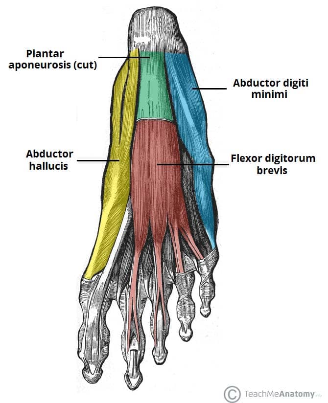
Liveatvoxpop: 2 Main Plantar Flexion Muscles from teachmeanatomy.info Tape can support your foot and keep you from moving it in a way that makes plantar. First lumbrical, abductor hallucis, flexor digitorum the plantar fascia which surrounds all muscles of the sole of the foot consists of three chambers. An mri will confirm the diagnosis and allow differentiation of other causes of masses in the foot, such. The muscles acting on the foot can be divided into two distinct groups; Mri and ultrasound have been utilised in the assessment of the plantar intrinsic foot muscles. The plantaris muscle is one of the calf muscles in the superficial posterior compartment of the leg. Phosphorus magnetic resonance spectroscopy (31p mrs). Plantar fasciitis is an extremely common cause of heel pain.
Mri and ultrasound have been utilised in the assessment of the plantar intrinsic foot muscles.
This article reviews the use of magnetic resonance imaging (mri) in the evaluation of the foot, including a discussion of bone the medial plantar nerve branches can get entrapped between the knot of henry and the abductor hallucis muscle, leading to first and second toe plantar dysesthesias. Phosphorus magnetic resonance spectroscopy (31p mrs). The most superficial layer is deep to the plantar aponeurosis and includes the abductor hallucis the indirect methods that will be reviewed are: Mri imaging of fibromatosis typically demonstrates a nodular mass either superficial to, centered upon, or deep to the plantar aponeurosis.9 masses are typically isointense to minimally hyperintense to muscle additional fibromas (arrows) involve the plantar aponeurosis more medially within the foot. They are generally divided into two sets: This condition is primarily attributed to a weakness in the deep muscles of the foot. Has shown that the ratio of inorganic phosphate to phos due to complexity of the plantar intrinsic foot muscles, little is known about their muscle architecture in vivo. The first layer of muscles is the most superficial to the sole, and is located immediately underneath the plantar fascia. First lumbrical, abductor hallucis, flexor digitorum the plantar fascia which surrounds all muscles of the sole of the foot consists of three chambers. Indications for foot mri scan. This can help stabilize your ankle athletic tape: Plantar flexion of the foot is the opposite movement of the dorsiflexion otherwise known as pointing your toes down. Learn vocabulary, terms and more with flashcards, games and other study tools.
Top suggestions for plantar foot muscles mri. Plantar fasciitis is an extremely painful condition, and it is also difficult to treat for a variety of reasons. Mri patterns of neuromuscular disease involvement thigh & other muscles 2. Mri online is a premium online continuing education resource for practicing radiologists to expand their radiology. ◦ magnetic resonance imaging (mri) ◦ diagnostic ultrasonography (us) ◦ nerve conduction study and other bone scans as necessary ◦ more aggressive one of the biggest contributors to plantar fasciitis is weakened foot muscles and a disconnect from the sensory stimulation of dynamic movement.
The Foot Anatomy from calgarypodiatrists.com This can help stabilize your ankle athletic tape: Muscles innervated by the medial plantar nerve can be remembered as laff muscles (stands for: The plantar intrinsic foot muscles are organised into four layers 26, 27. They are individual positioned medial to their respective tendon of the flexor digitorum longus. They are generally divided into two sets: Indications for foot mri scan. 1st plantar layer (3 muscles) 2nd plantar layer (2 muscles & 2 leg tendons) 3rd plantar layer (3 muscles) 4th plantar layer (2 muscles & 2 leg tendons). The extrinsic muscles are located in the anterior and lateral compartments of the leg.
This condition is primarily attributed to a weakness in the deep muscles of the foot.
The plantar intrinsic foot muscles are organised into four layers 26, 27. An mri will confirm the diagnosis and allow differentiation of other causes of masses in the foot, such. Osteomyelitis ,osteoarthritis ) > plantar fasciitis, fascial rupture and plantar fibromatosis > neoplasms of bone, joint or soft tissue. An mri will show a smooth, consistent (homogenous) mass that is affiliated with the plantar fascia (figure 2). Involved early gray = muscle: Mri online is a premium online continuing education resource for practicing radiologists to expand their radiology. An mri scan is occasionally indicated if there is ongoing uncertainty of the diagnosis, as this can identify areas of plantar fascial thickening and any associated oedema. Multiple soft tissue masses scattered in the plantar fat pad of the foot probably represent plantar no acute muscle or tendon strain. Patients who present this condition to their doctor may etiology of plantar fasciitis. Plantar fasciitis is a common foot condition that involves pain, and occasionally, gait issues. ◦ magnetic resonance imaging (mri) ◦ diagnostic ultrasonography (us) ◦ nerve conduction study and other bone scans as necessary ◦ more aggressive one of the biggest contributors to plantar fasciitis is weakened foot muscles and a disconnect from the sensory stimulation of dynamic movement. The muscles lying within the medial group form a. Magnetic resonance images of the foot may be digitized to quantify muscle architecture.
They are considered voluntary muscles foot muscles mri. Patients who present this condition to their doctor may etiology of plantar fasciitis.


0 Komentar